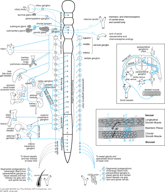
Schematic representation of the autonomic nerves and effector organs based on chemical mediation of nerve impulses. Blue, cholinergic; gray, adrenergic; dotted blue, visceral afferent; solid lines, preganglionic; broken lines, postganglionic. In the upper rectangle at the right are shown the finer details of the ramifications of adrenergic fibers at any one segment of the spinal cord, the path of the visceral afferent nerves, the cholinergic nature of somatic motor nerves to skeletal muscle, and the presumed cholinergic nature of the vasodilator fibers in the dorsal roots of the spinal nerves. The asterisk (*) indicates that it is not known whether these vasodilator fibers are motor or sensory or where their cell bodies are situated. In the lower rectangle on the right, vagal preganglionic (solid blue) nerves from the brainstem synapse on both excitatory and inhibitory neurons found in the myenteric plexus. A synapse with a postganglionic cholinergic neuron (blue with varicosities) gives rise to excitation, whereas synapses with purinergic, peptide (VIP), or an NO-generating neuron (black with varicosities) lead to relaxation. Sensory nerves (dotted blue lines) originating primarily in the mucosal layer send afferent signals to the CNS but often branch and synapse with ganglia in the plexus. Their transmitter is substance P or other tachykinins. Other interneurons (white) contain serotonin and will modulate intrinsic activity through synapses with other neurons eliciting excitation or relaxation (black). Cholinergic, adrenergic, and some peptidergic neurons pass through the circular smooth muscle to synapse in the submucosal plexus or terminate in the mucosal layer, where their transmitter may stimulate or inhibit gastrointestinal secretion.